

SCAPULA (OX) – LONG TYPE QUESTIONS-1st Year B.V.Sc & A.H (Veterinary Anatomy)
- The scapula is classified as a flat bone in anatomy.
- Present on the cranial part of the lateral thoracic wall.
- It is oriented in a downward and forward direction.
- The long axis of the scapula runs obliquely from the 3rd to 4th thoracic spine to the ventral (distal) extremity of the first rib.
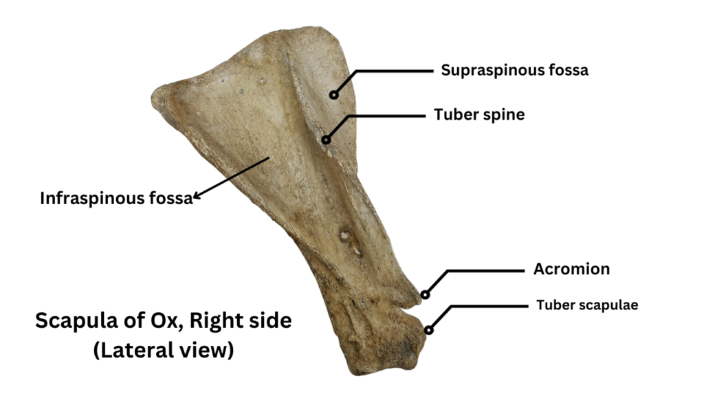
- Scapula is triangular in shape.
- Displaying a triangular outline, it features two surfaces, three borders, and three angles.
Surfaces of Scapula (Lateral & Medial/Costal)
1. Lateral surface
- The lateral surface of the scapula is divided into two fossae by its spine.
- The supraspinous fossa is positioned cranially to the spine, while the infraspinous fossa is located caudally.
- A rudimentary prominence (tuber), known as the tuberosity of the spine (Tuber spine), is present at its midpoint, serving as the attachment site for the trapezius muscle.
- The spine extends further into a pointed projection known as the acromion, which serves as the origin point for a portion of the deltoideus muscle.
- Supraspinous fossa:
- The supraspinous fossa, the smaller of the two, is smooth and houses the supraspinatus muscle.
- Infraspinous fossa:
- The infraspinous fossa accommodates the infraspinatus muscle.
- 2. Medial surface / Costal surface:
- The medial surface features a subscapular fossa (shallow) that runs its length.
- Subscapular fossa:
- The subscapular fossa occupies the majority of the ventral part of the surface, providing space for the subscapularis muscle.
- Dorsally , it separates two rough triangular areas, facies serrata, where the serratus ventralis muscle attaches.
- The cranial triangular area is designated for the attachment of the serratus cervicis muscle.
- The caudal triangular area is intended for the attachment of the serratus thoracis muscle.

- Borders of scapula (cranial, caudal, dorsal)
- 1. Cranial border:
- The cranial border is thin and convex.
- 2. Caudal border:
- The caudal border is thick and convex.
- The nutrient foramen is typically located in the ventral third of the caudal border.
- 3. Dorsal border:
- The dorsal border carries the scapular cartilage.
- The cartilage, an unossified part of the fetal scapula, fits into these depressions and elevations.

- Angles of scapula (cranial, caudal, ventral):
- 1. Cranial angle:
- The cranial angle is located at the junction of the cranial and dorsal borders.
- It is relatively thin and approaches a right angle.
- 2. Caudal angle:
- The caudal angle is thick and rough.
- It is formed by dorsal border and caudal border.
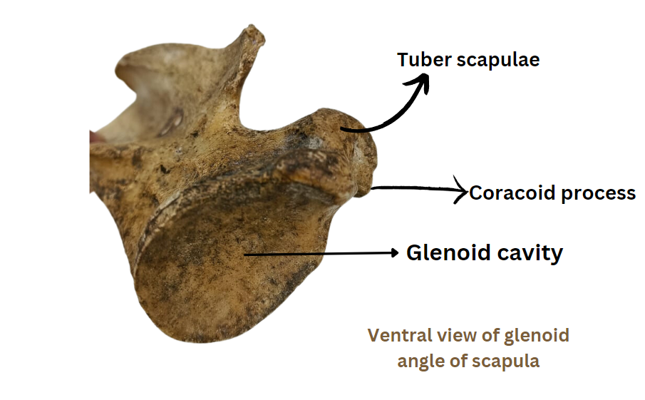
- 3. Ventral angle:
- It bears glenoid cavity and supraglenoid tubercle (tuber scapulae).
- In oxen, the glenoid cavity is nearly circular without a distinct notch.
- Glenoid cavity articulates with head of humerus.
- The supraglenoid tubercle (tuber scapulae) in oxen is small and close to the glenoid cavity.
- The tendon of origin of the biceps brachii muscles arises from tuber scapulae.
- The short and rounded coracoid process, from which the coracobrachialis muscle arises, projects from the medial side of the tuber scapulae.

